Cryo-electron microscopy (Cryo-EM)
- The cryo-EM suite at the Department of Structural Biology, located in the basement of BST3, is equipped with state-of-the-art electron microscopes for high-resolution three-dimensional structural analysis of protein assemblies and cellular components.
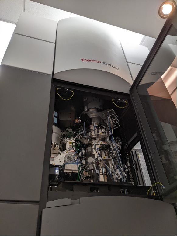
- The roster includes four Thermo Fisher (previously FEI) microscopes – a Titan Krios 3Gi equipped with a Selectris energy filter and a Falcon 4i direct electron detector; a TF20 equipped with a TVIPS XF416 camera and Gatan cryoholders; a T12 equipped with a Gatan Orius CCD camera; and an Aquilos 2 focused ion beam (FIB) mill for thinning cellular samples. The Krios and TF20 are capable of low-dose “single-particle” and tomographic studies while the T12 is used for optimizing samples by negative-stain EM. The facility is also equipped with a FEI Vitrobot Mk4 for frozen-hydrated specimen preparation and other ancillary equipment for cryo-EM.
- Conventional negative-stain electron microscopy provides two-dimensional (2D) projection images of the stain-embedded specimen in which the distribution of the heavy-metal stain represents the stain-excluding shape of the specimen. Cryo-EM offers a more direct representation of protein structure that avoids the dehydration and chemical modification of classical methods by immobilizing the specimen in a thin layer of vitrified (non-crystalline) buffer.
Single particle cryo-EM
- Biological specimens are radiation sensitive, requiring very low-intensity images that have very noisy backgrounds. To overcome this problem, images recorded from thousands of identical copies of specimens randomly oriented relative to the electron beam are combined and averaged. This method is referred as “single particle” microscopy as the particles are “free-standing” or “single” as opposed to participating in a higher level of assembly such as a crystal. Many structures have been determined by cryo-EM, recently to atomic resolution, and ranging in size from 50kDa to 200MDa. These structures reveal tertiary, secondary and primary structural elements as well as interfaces between subunits, and usually in a native, functional context. CryoEM is applicable for protein complexes that are too large and/or too heterogeneous to be investigated by high resolution X-ray and NMR methods.
Cryo-electron tomography
- Electron tomography (ET) is used to solve structures of “one-of-a-kind” objects for which averaging methods cannot be applied, such as cellular components like organelles. The underlining principle is similar to computerized tomography (CAT scans) where the specimen is rotated relative to the incident electron beam and a series of projection images are combined computationally to reconstruct the 3D structure of the object. This method can be applied to the thinnest regions of cells (typically <0.4 µm thick) or to whole cells that are thinned by FIB-milling, and yields valuable “in situ” information on the structure, organization and distribution of proteins, membranes and organelles.
Visit the CryoEM Center page here